Is a Patient With a Concussion Admitted to the Hospital?

Recently, I finished a book that included the following medical scenario. The main character fell into a river and suffered a broken arm and concussion. During her ER visit, the doctor tells her she needs to be admitted overnight for observation because of the concussion.

This is a common medical myth (along with the one that a CT scan is required in all instances of head injury– it’s not.)

A simple concussion does not need an overnight hospital stay. Let me qualify what I mean by simple. You receive a hit on the head and have one or some of the following global symptoms (dizziness, headache, nausea, vomiting, and amnesia to the events.) Global symptoms mean more than just the bump on your head hurts.

This is really how concussion is diagnosed. CT scan is reserved for concerns of bleeding and/or fracture that might require a neurosurgical intervention. Typically, symptoms associated with bleeding and fracture are persistent and more dramatic. Headache pain is not relieved with medication and/or worsens. There is more than one episode of vomiting. Persistent confusion. Perseverating– saying the same thing over and over. Inability to move part of the body. Decreased responsiveness. Amnesia that doesn’t improve.

A patient with a simple concussion is monitored in the ER for several hours. Typically, we’ll give them medication based on their symptoms to see if they improve. For instance, a patient that has nausea, headache and dizziness will get an anti-nausea medication and an over-the-counter pain reliever like Tylenol or Ibuprofen. If their symptoms improve and/or resolve and they can hold something down to eat then they are discharged home with instructions on when to return to the ER.

In order to be admitted into the hospital the patient must exhibit severe, persistent symptomology and/or have bleeding and/or fracture.

In absence of these, the patient will be discharged home.

EMS and ER Response for an Unconscious Female Trauma Patient


Ginger Asks
:

I have a 23-year-old woman with an obvious head wound (she got hit with the butt of a gun, but the first responders don’t know that) who’s been outside in 20’ish degree weather without a coat for an undetermined amount of time. She’s unconscious. Obviously an IV is started, but what else will paramedics do to treat her? Warming blankets? What would happen when she got to the ER?

Jordyn Says:

Thanks for sending me your question.
 
EMS Response:

For an unconscious patient with an obvious head wound, but is unable to tell how her injury happened should be placed in C-spine precautions. That means C-collar and backboard. IV– yes. And warming. They’d get a set of vital signs, put her on a monitor and then do a full assessment to look for other injuries.

Checking her blood sugar is warranted because why is she unconscious? Did the injury to her head happen because she passed out from low blood sugar? Or is it too high? Looking for medical alert bracelets as well. They’d probably key in on a good neuro exam like are her pupils equal and reactive to light? What type of stimulation does she respond to (voice, touch or pain?) They might even give a dose of Narcan to rule out opiate overdose (like heroin.)  


In the ER:

Full assessment as above and we’ll look for other injures. We’ll maintain C-spine precautions. She would be completely undressed (again– looking for other injuries.) We have a better ability to monitor temperature so we’ll know exactly where she’s at and work to rewarm her. This could range from warm blankets to warming lights and heated IV fluids. Full set of vital signs. We’d place her on the monitor as well to watch her HR, breathing and oxygen levels continuously.
 
As far as testing and procedures go, if she remains unconscious, I would say the following:

1. Spine X-rays. 
2. CT of the head (to look for bleeding, stroke, tumor.)
3. Labs: Full metabolic panel (this will check blood sugar again), complete blood counts, alcohol level, aspirin level, Tylenol level. Tylenol and aspirin are drugs people will overdose on that can be very serious.
4. Urine toxicology panel (this would pick up on major substances of abuse but not everything.) Also urine pregnancy test. 
5. ECG. To see if a heart arrhythmia or heart attack could be an explanation for her passing out.
 
Unless we know the exact mechanism of the injury we have to consider both inflicted wounds from another person but also that she might have just passed out and hit her head and what the reason for that might be.

If she’s truly unconscious and doesn’t respond to pain– she’d likely get a tube in every orifice as they say and they’d have to consider whether or not to intubate her (put a breathing tube in) to protect her airway. If that happens, then NG tube (placed probably through the mouth into the stomach) and a Foley catheter which drains your urine into a bag.

If she’s somewhat responsive but immediately drifts off– they could hold off on tube placements, check the tests I’ve listed, and give her some time to see if she wakes up on her own if she’s breathing well on her own.

Head Injuries: Jason Joyner

There was that time when the editor saved the medical professional.

As a physician assistant, I enjoy having medical aspects in my story. But even medical folks can slip up and have errors in our fiction.

I have a scene where my heroine gets head trauma and wakes up later in the clutches of the villain. The freelance editor, Ben Wolf, wondered about that. He had read that if there was significant time of loss of consciousness (LOC), then it suggested a serious injury that would be hard for the victim to bounce right back from to be active.

One of my pet peeves is when characters are injured and recover too fast, so I had to look into this again.

Basically, my heroine suffered a concussion, also known as a Traumatic Brain Injury (TBI). Symptoms of a concussion can include headache, confusion, dizziness, visual changes, a blunted affect, and may or may not include LOC. (People always flash lights in pupils to check for concussion. If the pupils are affected, it is a serious sign and they won’t be up and active soon.)

LOC usually is only for a few minutes, and as my editor noted, will mean a much more severe injury if it lasts for hours.

Blast. Foiled by the editor.

Except, you can use the amnesia angle.

A concussion with LOC may have retrograde (before the incident) or antegrade (after the incident) amnesia. According to one research article, the antegrade amnesia can last for a few hours after the incident. I can attest – I had a concussion in 5th grade and couldn’t remember a couple hours afterwards.

So if you need your protagonist to be out of it for a while, keep the actual LOC on the short side and use the amnesia angle to get you where you need to be. The victim may be incoherent, unsteady, with a blank expression during this time. Use these symptoms to add drama to the situation.

When your protagonist comes to, it is actually the end of antegrade amnesia. I remember with my concussion it was like I “woke up” after lunch during our quiet reading time at school. I was confused, unsure of what happened. I could remember part of the morning, but about two hours was blank. I even found a goose egg on my head later, but I didn’t know how it got there.

So that was my work around. My heroine didn’t have LOC the whole time. But there was enough injury to cause confusion and amnesia, keeping her from attempting escape. There you go Ben. A few minor tweaks, and all is well. Except for my heroine, who’s tied up and threatened. But that’s another story.

**************************************************************************

Jason loves good stories and wants to use words to make a difference. When he’s not writing, playing soccer, or losing in fantasy football, he works as a physician assistant in southeast Idaho. He also tries to keep up with his awesome wife, three high-energy boys, and his little princess. He writes suspense and YA supernatural, and likes to use his medical experience to punch up the stories. You can find him on Twitter @JasonCJoyner or his blog at www.jasoncjoyner.com/blog.

Dianna T. Benson: A Son’s Tale of Traumatic Brain Injury


The term concussion is well known. The medical field refers to a concussion as a TBI – Traumatic Brain Injury. Contact sports are one of the top causes of a TBI, another are MVCs – Motor Vehicle Collisions.
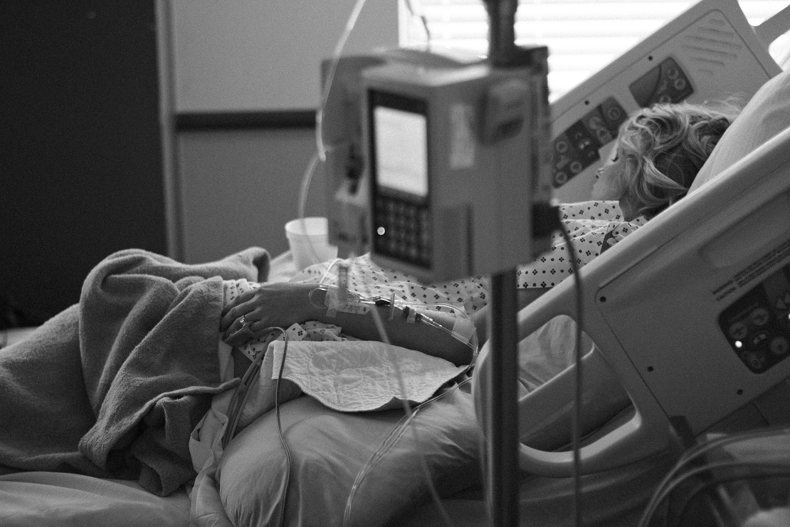
My teenaged son has endured four concussions. The first two as a goalie for the Junior Hurricanes and the third in a MVC. The first one took him out of school for a month and hockey for three months. The second, a year later, was more mild, which is unusual. Typically, a patient suffers a more severe TBI the second time. In the MVC, a classmate was driving them to school when another car struck them. This third TBI ended my son’s hockey career, preventing him from attending the Junior Hockey draft in Canada Spring of 2013.
The problem wasn’t simply that this was his third concussion, although that in itself is a strong reason to end a contact sport career. With this third TBI, a neurologist evaluated him versus just the concussion clinic MDs who’d treated him with the first two. Not only was it his third TBI, but his symptoms were extremely severe, which didn’t make sense to me – the details of the MVC didn’t suggest such injuries for my son: 1) None of the others involved in the crash suffered any injuries 2) No air bags deployed 3) Vehicle damage was minor. As an EMT for nearly a decade, I wondered about underlining health conditions in my son. I also considered he had not fully recovered from the first two concussions and was in denial about his symptoms in order to play hockey.
Sure enough, the neurologist diagnosed my son with hyper-mobile joints (something I already knew but wasn’t aware of the danger with contact sports.) The MD also diagnosed him with mild CP (cerebral palsy), a diagnosis that made sense to me since my son was born in respiratory arrest and was non-verbal and had spasticity until over age two. Both diagnosis are a recipe for injury, especially in contact sports. The MD gently told my son he was done playing goalie forever – it was devastating and crushed him. Understanding his hockey career was over, he admitted he’d ignored symptoms because he had a shot to play Junior Hockey, college hockey, and possibly professional hockey. A life-long athletic competitor myself, I completely understood the denial that led him to ignore his body.
Hyper-mobile joints, while creating an incredibly athletic body, are highly susceptible to any musculoskeletal injury in that individual. For my son, after two TBIs in a contact sport, his hyper-mobile neck was easily and severely whip-lashed in the MVC, jostling his brain fiercely, causing all his concussion symptoms to return and more heightened than ever.
Ten months after the car accident, the fourth TBI occurred December 2013 just days after the neurologist cleared my son to return to his life minus contact sports. The neurologist gave my son the green light to snowboard. That December day on the mountain, my son didn’t even hit his head and he sustained no head trauma – simply snowboarding jostled his brain enough to cause another TBI.   
Even though he’s extremely athletic, my son’s body shouldn’t do what it can to do. The risk of permanent brain damage and partial or full paralysis is too high for him– something he now understands. I described it to him as this: When Cam Ward (the goalie for the NHL team Carolina Hurricanes) is playing goalie, his body is naturally like a SUV of protection in a MVC. Whereas, for my son, his body is like a motorcycle in a MVC – no protection.
Until Spring 2015, my son is restricted from doing anything with speed, wheels, height or repetition (basically everything fun.) This next year his brain will heal, then little by little he can attempt things (no contact sports ever, though) to see how his body responds. At 6’7” in height and extremely athletic, he appears a medically sound seventeen-year-old, but inside his body tells a different story. 


God works in amazing ways and this is my son’s blessing. Since cerebral palsy only affects motor function, and none of the four TBIs caused him any loss of cognitive abilities, he’s still as annoyingly brilliant as ever and is anxious to head off to college this fall. For now, his goal is to graduate medical school with a degree in neurology and become a neurosurgeon since he feels (understandably so) he can relate to patients’ symptoms with head trauma. 
***********************************************************************


 Dianna Torscher Benson is a 2014 Selah Award Finalist (winners not yet announced), a 2011 Genesis Winner, a 2011 Genesis double Semi-Finalist, a 2010 Daphne de Maurier Finalist, and a 2007 Golden Palm Finalist. In 2012, she signed a nine-book contract with Ellechor Publishing House. She’s the author of The Hidden Son, her debut novel. Final Trimester is her second release.
After majoring in communications and a ten-year career as a travel agent, Dianna left the travel industry to earn her EMS degree. An EMT and a Haz-Mat and FEMA Operative since 2005, she loves the adrenaline rush of responding to medical emergencies and helping people in need.
Dianna lives in North Carolina with her husband and their three children. 
Her releases are available wherever books are sold. Below are the links to Final Trimester at the three largest booksellers:


Author Question: Speech Therapy after Traumatic Brain Injury

Karen Asks:

I’m writing a story about a man who is shot in the head in a way that impacts his ability to speak.  Long months of rehab restore his speech but leave him with a stutter.  Is this feasible?  Which part of the head would he need to be shot in?  What else could be impacted by such a wound?  Can you recommend any websites or resources about gunshot wounds or speech therapy?

Jordyn Says:

Karen– thanks so much for sending me your question.

Generally, the left side of the head is considered to contain the speech centers of the brain–in most cases. It might depend on whether or not your character is right or left handed.

97% of right handed people have their speech centers on the left hemisphere.

19% of left handed people have their speech centers on the right hemisphere– which may be where the phrase “left-handed people are the only ones in their right mind” come from. I LOVE this phrase speaking as a left-handed person.

68% of people have language abilities in BOTH hemisphere.

To read more on these areas– check out this link: http://webspace.ship.edu/cgboer/speechbrain.html
A good case to look into would be former Arizona Congresswoman Gabrielle Gifford’s. She received a serious gunshot wound to the head and had extensive rehab– over many many months. It might give you an idea of how long the road to recover is for some of these victims. It can be years.

http://www1.uabhealth.org/BoneandJoint/victims-gunshot-head-daunting-road-ahead

http://www.aans.org/en/Patient%20Information/Conditions%20and%20Treatments/Gunshot%20Wound%20Head%20Trauma.aspx

http://www.everydayhealth.com/blog/dr-black-brain-health/the-outlook-for-recovery-from-a-gunshot-wound-to-the-brain/

I think you have a lot of leeway as an author to decide what you want to do after brain injury because we don’t understand as much about the brain as we do other organs. It might be hard to pinpoint sources of “speech therapy after gunshot wound to the head” (which is how I first started to Google your inquiry) but a gunshot wound would be considered a traumatic brain injury so I started to Google that and came up with several other resources as well. Here’s a great You Tube Video that demonstrated a speech therapy session that could be great for a fiction novel.

What else could be impacted? Anything really. Again– you have a lot of leeway here. There could be motor issues as well. Difficulty walking. Difficulty with fine motor skills. To the other extreme which would be coma.

***************************************************************************

After creating Christian education curriculum for 25 years and writing over 250 published articles, Karen Wingate has turned her attention toward historical and contemporary fiction.  She lives with her husband and Welsh Corgi in Western Illinois.

Author Question: Death by Trophy

Susan Asks:

I have a woman murdered when she is hit on the back of the head with a metal trophy. The trophy is cup shaped so the largest part of it is a thinner metal. I expect the trophy will dent from the impact, but I’d also expect that there would be blood as a result of the injury. Would this kind of injury cause bleeding and if so can you give me a general idea of how much?

Jordyn Says:

It depends. Blows to the head can go either way. They can just cause internal bleeding (intracranial hemorrhage) and/or an external scalp laceration that would bleed A LOT depending on it’s size and depth. Scalp wounds are known for being pretty bloody.

These injuries can be nice for your character as you have some leeway medically to do them in as you please. 

Why McDreamy is the Worst Neurosurgeon Ever

First and foremost, let me say that I am a Grey’s Anatomy fan– not for the medical accuracy for sure– but it is a guilty pleasure of mine. So, considering the known medical inaccuracies I’ve seen, I don’t have to be a neurosurgeon to guess that they are likely a little loose with the surgical details.

But this one I could not ignore.

Neurosurgeons place VP (ventriculostomy-peritoneal) shunts. What is that?

A VP shunt is a tube that is placed in a person’s brain, specifically the ventricle, to drain off excess cerebrospinal fluid (CSF). Placing a VP shunt is primary treatment for a condition called hydrocephalus where there is excess accumulation of CSF in the brain. Too much CSF will lead to increased intracranail pressure– which can be deadly.

A VP shunt helps keep the brain at an even pressure by draining extra fluid into the perotoneal cavity (or your gut.)

Here is a primer on traumatic brain injury. This post covers some important principles of managing traumatic brain injury. For quick review, the skull contains three components: your brain, blood, and CSF.

Back to Grey’s.

Derek (aka McDreamy) and his surgeon wife adopt a baby named Zoila. One of the principle reasons for the adoption was that the child (approx 2 y/o) had a VP shunt and Dr. McDreamy would be an expert at managing her condition.

Really?

So, when the child begins to have vomiting and fever– he makes the statement, “Well, she just has the flu.”

The first thought in a neurosurgeon’s mind, until ruled otherwise, is that something is wrong with the shunt in her head!

This concept is drilled into emergency medical personnel– particularly pediatrics, that if a person with a VP shunt presents with headache, vomiting, and fever– it is an emergency. First assumption is something is wrong with the shunt and if not treated, the person could have elevated intracranial pressure (which is bad) and die (which is super bad!)

Classic symptoms for increased pressure in the brain is headache and vomiting. These could mean that the shunt is obstructed and no longer draining CSF. Fever could indicate the shunt is infected– which essentially means the patient has a brain infection (meningitis)– another really bad thing.

So for super surgeon, McDreamy, to blow this off as a viral illness is a big no-no. Off to the ER little Zoila should have gone.

Determining Brain Death: 3/3

Last post, we talked about the use of apnea testing to determine brain death after the patient meets certain criteria.

There is one additional test that may be done to determine brain death and that is a brain perfusion scan.

This procedure is done in radiology which can make it very difficult. Imagine taking a ventilated patient through the halls of the hospital along with several IV pumps giving medication that are keeping the patient alive. That in itself is not a fun excursion.

Once the patient is in radiology, they are given an injection of a radioisotope—something that will trace where the blood is flowing. After the injection, photos are taken of the patient’s brain. If there is no blood flow to the brain, and this must include the brain stem as well, then the patient is said to have “brain death” and is clinically dead at that point.
This You Tube video provides a very good explanation of these concepts.

After brain death is determined, the patient is not immediately withdrawn from life support but a conversation will ensue with the family that the patient has died and they will be encouraged to discontinue life support.

Generally, families are given a lot of time to come to terms with this decision. Anywhere from 1-3 days is reasonable. They may want to fly in additional family members to be present when life support is discontinued. I’ve never been part of a situation where, when the finding of brain death were fully explained, where families chose not to discontinue support.

This is not to say that the patient may not proceed to circulatory death despite receiving life support. Once the brain has died, it does become very difficult to keep the body functioning.

Does this change your mind about how brain death is determined?

Determining Brain Death: 2/3

I’m continuing with a series on how brain death is determined. All hospitals likely have a policy in place with strict guidelines on how brain death is determined. Check last post for the beginning stages.

Now, we’ll move onto actual testing.

Can the patient breathe on their own? This is a relatively simple test. It’s called apnea testing. The ventilator is turned off and we see what the patient will do. Naturally, when we stop breathing, carbon dioxide will build up in the blood stream. Your body has receptors that monitor the level of CO2 and it will initiate a breath when the levels rise.

Here is the procedure for performing an apnea test.

1. The patient will be on an ECG and pulse ox monitor.

2. Give the patient 100% oxygen for five minutes.

3. After five minutes, disconnect the patient from the vent, but give oxygen via T-piece. The breathing tube will still be in place. At this point, the patient is off the vent and no longer being assisted but will have needed oxygen if they do initiate a breath.

4. Watch the patient for breathing. If any attempt is made to breathe, it is inconsistent with brain death and the test is stopped and the patient is placed back on the ventilator.

5. If the patient has any cardiac arrhythmias, low blood pressure or oxygen level that falls to less than 80% (normal level is 90-100%) then the test is discontinued. These finding will lead more to a conclusion that brain death has occurred.

6. If the carbon dioxide level increases above 60 (normal level is 35-45)—the apnea test is consistent with brain death. The brain is very sensitive to rising levels of carbon dioxide and the absence of a response is consistent with brain death.
Next post, we’ll talk about brain perfusion studies.

Determining Brain Death: 1/3

Several months ago, I skewered a Hallmark movie for its unrealistic portrayal of discontinuing life support. In light of that, I thought I’d do a special series on determining brain death.


How do medical personnel determine a patient has suffered brain death?

Brain death means that your brain as an organ has died. It is no longer receiving blood flow. Without blood flow, no oxygen is being delivered. Without oxygen, an organ dies. Your brain is your body’s main control. If it has died, you have died.

If you have a character that is brain dead, they should be on life support. Again, if the brain isn’t working, it’s not telling your lungs to inhale. However, we can do this medically with a ventilator. This is why families sometimes have trouble understanding brain death means ultimate death. If we provide oxygen to the lungs, the heart will continue to beat and bodily functions can be maintained for a limited amount of time. A family sees the rise and fall of the patient’s chest and assume the patient is initiating those breaths when in fact it is the machine doing all the work.


There are several ways to determine brain death. Some are not as precise as others. I’ll try to cover least precise to most precise.


Before testing, there is generally an observation period. My hospital uses the following guidelines:


Less than 7 days: Not applicable
Age 7 days-2 months: 2 exams 48 hours apart
Age 2-12 months: 2 exams 24 hours apart
Over 12 months: 2 exams 12 hours apart
Adults (18 years and older): 2 exams 6-12 hours apart.


Also, prior to the exam to determine brain death, the patient must also meet the following criteria:


1. Absence of a reversible condition. The cause of the coma must be documented.


2. Absence of hypothermia. The patient must have normal body temperature.


3. Absence of hypotension. The patient must have normal blood pressure.


4. Absence of drugs or toxins in significant amounts as to interfere with the diagnosis of brain death.


5. Absence of a metabolic cause of the coma.


6. Normal levels of carbon dioxide.


Once these are met, the patient should be observed for the following:


1. No cranial nerve reflexes. Here is an extensive list of what those are: http://www.clinicalexam.com/pda/n_cranial_nerves_exam.htm


2. Flaccid tone in all extremities.


3. No response to deep pain.


Once these are met, the patient proceeds to apnea testing. That’s where we’ll pick up next post.